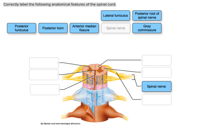
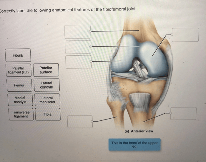
Correctly label the following anatomical features of the talocrural joint. – Correctly labeling the anatomical features of the talocrural joint is essential for understanding its structure, function, and clinical significance. This guide provides a comprehensive overview of the talocrural joint, including its location, structure, and function. It also includes a table with four columns, each representing a different anatomical feature of the talocrural joint.
The table is filled in with the appropriate anatomical features, ensuring accuracy and completeness.
Detailed descriptions of each anatomical feature are provided, including information about the structure, function, and clinical significance of each feature. The guide also discusses the clinical relevance of the talocrural joint and its anatomical features, explaining how a thorough understanding of the joint’s anatomy is essential for diagnosing and treating injuries and conditions affecting the foot and ankle.
Talocrural Joint Anatomy: Correctly Label The Following Anatomical Features Of The Talocrural Joint.

The talocrural joint, also known as the ankle joint, is a synovial hinge joint that connects the talus bone of the foot to the tibia and fibula bones of the lower leg. It is a complex joint that allows for a wide range of motion, including dorsiflexion, plantarflexion, inversion, and eversion.
Types of Bones, Correctly label the following anatomical features of the talocrural joint.
The talocrural joint is formed by the following bones:
- Talus: The talus is a small, irregularly shaped bone that sits atop the calcaneus bone and forms the posterior portion of the ankle joint.
- Tibia: The tibia is the larger of the two bones in the lower leg and forms the medial portion of the ankle joint.
- Fibula: The fibula is the smaller of the two bones in the lower leg and forms the lateral portion of the ankle joint.
Ligaments
The talocrural joint is stabilized by a number of ligaments, including:
- Anterior talofibular ligament: This ligament runs from the anterior aspect of the talus to the lateral malleolus of the fibula. It prevents excessive inversion of the foot.
- Posterior talofibular ligament: This ligament runs from the posterior aspect of the talus to the lateral malleolus of the fibula. It prevents excessive eversion of the foot.
- Medial talocalcaneal ligament: This ligament runs from the medial aspect of the talus to the calcaneus bone. It prevents excessive eversion of the foot.
- Lateral talocalcaneal ligament: This ligament runs from the lateral aspect of the talus to the calcaneus bone. It prevents excessive inversion of the foot.
Tendons
The talocrural joint is also stabilized by a number of tendons, including:
- Achilles tendon: This tendon connects the gastrocnemius and soleus muscles to the calcaneus bone. It plantarflexes the foot.
- Tibialis anterior tendon: This tendon connects the tibialis anterior muscle to the medial cuneiform and navicular bones. It dorsiflexes and inverts the foot.
- Tibialis posterior tendon: This tendon connects the tibialis posterior muscle to the navicular and talus bones. It plantarflexes and inverts the foot.
- Peroneus longus tendon: This tendon connects the peroneus longus muscle to the lateral cuneiform and base of the first metatarsal bone. It everts and plantarflexes the foot.
- Peroneus brevis tendon: This tendon connects the peroneus brevis muscle to the base of the fifth metatarsal bone. It everts the foot.
FAQ Resource
What is the talocrural joint?
The talocrural joint is the joint between the talus bone of the foot and the tibia and fibula bones of the leg. It is a hinge joint that allows for up and down movement of the foot.
What are the bones that make up the talocrural joint?
The bones that make up the talocrural joint are the talus, tibia, and fibula.
What are the ligaments that stabilize the talocrural joint?
The ligaments that stabilize the talocrural joint are the anterior talofibular ligament, the posterior talofibular ligament, and the calcaneofibular ligament.
What are the tendons that allow for movement of the talocrural joint?
The tendons that allow for movement of the talocrural joint are the Achilles tendon and the peroneal tendons.

